baseline vaginal ultrasound
Maximum thickness of 29 cm was displayed by double layered measurement of the adherent vaginal walls. General Transvaginal ultrasound is preferred and usually mandatory modality for monitoring follicles.

Transvaginal Ultrasound Images Showing Typical Features Of A Right Download Scientific Diagram
Four surgeons performed operations on 85 patients.

. The purpose is to check that there are no unusual cysts on the ovaries before starting the fertility drugs. Summary of Key Points. Similarly vaginal pressure readings at.
The mean creatinine in patients with vesicovaginal fistulas VVF was 060 ngml versus patients with uretero-vaginal fistulas UVF 079 ngml P 0012. Ultrasound remains one of the most routinely performed medical investigations and a mainstay of clinical decision-making rests upon the obtained results. This isnt dangerous and will usually go away without intervention.
12 Sometimes a stubborn corpus luteum cyst sticks around even after your period starts. Find an intrauterine device IUD. The goal of this scan is to check how many antral follicles you have in each ovary and to ensure there are no abnormalities.
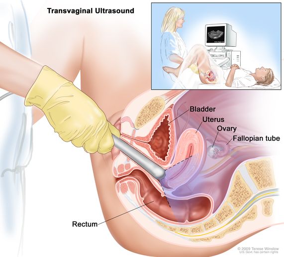
Baseline creatinine and renal ultrasound findings. If you are undergoing infertility treatment you will have a baseline ultrasound where we check to confirm that your ovaries have not formed any unexpected cysts that may disrupt your treatment. The sound waves bounce off the organs inside your body and the probe picks them up.
Normal anatomy of the vagina on transperineal ultrasound There are differences in vaginal dimensions among females. A self-administered baseline questionnaire completed before ultrasound screening provided information on sociodemographic characteristics age race marital status and education body weight and height reproductive and obstetric history family history of cancer personal medical history cigarette smoking and use of hormone replacement. Due to the relative proximity of the pelvic organs to the abdominal surface and easy access via the vaginal route gynaecological scanning should be in the armamentarium of every gynaecologist.
Transperineal ultrasound imaging was performed with a Toshiba SSA-340S ultrasound machine Toshiba Japan equipped with a 57-MHz endocavitary probe. Ultrasound monitoring may begin on day 3 of the cycle to assess a baseline size as well as exclude if any cyst remains from previous hyperstimulation or otherwise. Vaginal health index perineometry measurement of the vaginal wall thickness by ultrasound Doppler sonography of the vaginal walls vessels optical coherence tomography biopsy cytological and immunocytochemical methods.
In women doctors can use a pelvic ultrasound to. A simple cyst less than 3 cm in the ovary of a premenopausal patient is best termed a follicle and is a normal finding. Transvaginal ultrasound scan An ultrasound scan is a procedure that uses high frequency sound waves to create a picture of a part of the inside of your body.
Look for cancer in your ovaries uterus or bladder. The ultrasound scanner has a probe that gives off sound waves. A baseline ultrasound is a scan done early in your menstrual cycle before starting any fertility medications.
The probe looks a bit like a microphone. Iui infertility vaginalultrasoundWatch as I discuss what the baseline ultrasound process was like what the doctor is looking for and what to expect next. Find problems with the structure of your uterus or ovaries.
Although improvements were seen in VAS scores for dysmenorrhea nonmenstrual pelvic pain and dyspareunia there were no significant differences in ultrasound biometry at either rest Valsalva or on contraction when comparing postinjection measurements at 4 12 and 26 weeks with pre-injection baseline. To assess the condition of the vaginal walls before and after laser treatment the following methods will be used. The length of the vagina from external os to introitus ranged from approximately 41 to 95 cm.
This is known as your baseline ultrasound. The examination was performed with the patient in the lithotomy position and with a half-filled bladder. The probe was covered with a condom after application of a thin layer of ultrasound gel.
A simple cyst less than 1 cm in the ovary of a postmenopausal patient is considered inconsequential. We also look at the endometrial lining thickness to make sure it. Most ovarian masses are benign and have a typical sonographic appearance that allows accurate diagnosis.

Transvaginal Ultrasound Of The Rectosigmoid With Signs Of Infiltration Download Scientific Diagram

Transperineal And Endovaginal Ultrasound For Evaluating Suburethral Masses Comparison With Magnetic Resonance Imaging Okeahialam 2021 Ultrasound In Obstetrics Gynecology Wiley Online Library

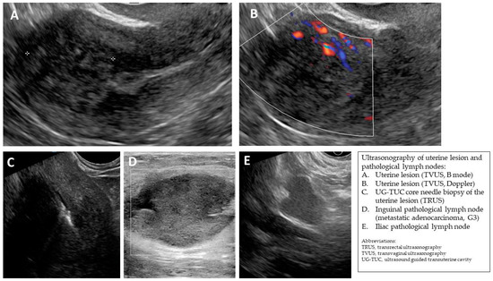
Jcm Free Full Text Transvaginal Ultrasound Combined With Strain Ratio Elastography For The Concomitant Diagnosis Of Uterine Fibroids And Adenomyosis A Pilot Study Html

Definition Of Transvaginal Ultrasound Nci Dictionary Of Cancer Terms Nci

Advanced Pelvic Ultrasound In House At Veritas Fertility Surgery
Gynaecology Ultrasound Women S Ultrasound Melbourne

Sonographic Characterization And Surveillance Of Paravaginal Smooth Muscle Tumor Of Uncertain Malignant Potential Zamora Journal Of Clinical Ultrasound Wiley Online Library

Transvaginal Ultrasound Images Showing Typical Features Of A Right Download Scientific Diagram
Gynaecology Ultrasound Women S Ultrasound Melbourne

Transvaginal Ultrasound Scan From A Patient With Spontaneous 46 Xx Download Scientific Diagram

Advanced Pelvic Ultrasound In House At Veritas Fertility Surgery

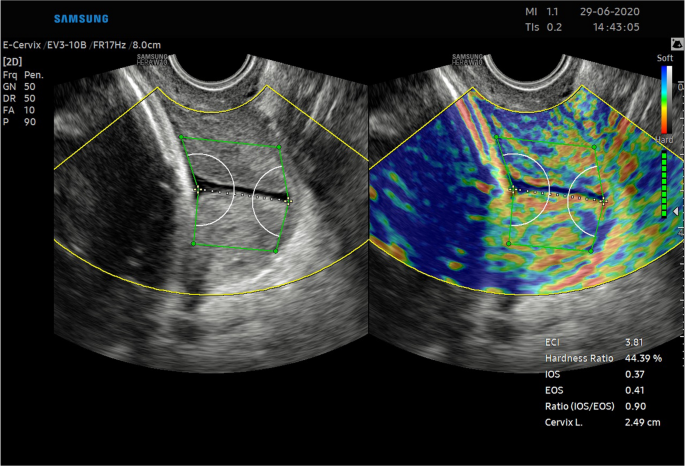
Repeatability And Reproducibility Of Quantitative Cervical Strain Elastography E Cervix In Pregnancy Scientific Reports

Transvaginal Ultrasound Depiction Of The Bladder A Normal Bladder Download Scientific Diagram

Diagnostics Special Issue Imaging Of Gynecological Disease

Transverse Ts And Longitudinal Ls Transvaginal Ultrasound Images Of Download Scientific Diagram

Ultrasonographic Techniques And Clinical Applications Springerlink


Comments
Post a Comment